Pediatric Interventional Radiology Online Handbook
  • Home
  • Procedures
  • Protocols
  • Online Library
    • Pediatric IR Papers
    • Presentations and Webinars
    • IR Equipment and IFU
  • BCH IR Fellows Homepage
  • About Us

​Hip Arthrocentesis

Picture
X
Picture
X
Picture
X

Introduction

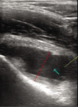
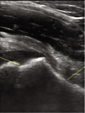
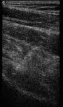
Hip septic arthritis is a surgical emergency. Prompt diagnosis and drainage are essential to prevent joint damage. In infants, septic arthritis may occur from propagation of adjacent osteomyelitis. Causative organisms include Staphylococcus aureus and Streptococcus pneumoniae. Pain is typically acute, localized, and severe (refuses to bear weight) and accompanied by fever. Although occasionally the presentation is more subtle or afebrile. Bilateral effusions suggest a systemic arthritic disorder or transient synovitis.

Guidelines

Guidelines for US-guided Hip Arthrocentesis
  • Prior to placing a request for an urgent US-guided hip arthrocentesis:
    • Sonographic confirmation of joint effusion. This study is performed by the ultrasound division during working hours or by the Pediatric Radiology fellow during nights and weekends.
    • Clear indication based on criteria for child with painful hip (see below)
  • IR team can be reached via IR NP Pager # 0434 during working hours or the IR fellow during nights (5-7 AM) and weekends.
  • The procedure is performed in the ED by the IR fellow using a portable ultrasound machine and appropriate tools. After-hours hip taps will not entail paging an on-call tech or nurse into the hospital.
  • The IR attending on call will be the physician of record for hip taps after hours.  It is at the attending’s discretion whether support is provided by phone or in person.
  • The ED team will provide sedation for these procedures.
  • Representative labelled US images (pre-, intra- and post-procedural) are recorded.
  • IR fellow will return the portable US to the designated area in IR and plug in the power and US data cable.
  • Early the next morning, the IR techs will confirm transfer of images and provide a code for dictation purposes.
  • IR fellow will email the IR team with details and needs for coming to work late.  
  • On weekdays during working hours, hip taps can be performed either in IR, using IR sedation resources or in the ED.  Criteria for sedation in IR will follow guidelines used in IR for all cases
  • It is understood that hip taps will be performed without undue delay.  However, each case will be triaged by the IR staff, along with other active and pending studies.

​Kocher 4 criteria for child with painful hip:
  1. Non-weight-bearing on affect side
  2. ESR > 40 mm/hr
  3. Fever > 38.5 C
  4. WBC count of >12,000 /ml
​
  • Chance of septic arthritis with positive criteria: 4= 99, 3= 93%, 2 = 40, 1= 3%.​

Indications

For Urgent Hip Arthrocentesis
  • ​Intermediate probability (two positive criteria) of septic arthritis.

​Management approach
​

Number of criteria

probability

Approach

0

Very low

Observation only

1

Low


2

Intermediate

May be good candidates for US-guided aspiration

3-4

High

Aspiration in the operating room instead of image-guided needle aspiration given the greater likelihood that they will require surgical drainage.


Diagnosis of septic arthritis
  • True septic arthritis: Positive culture of synovial fluid or blood + fluid WBC > 50 × 109/L
  • Presumed septic arthritis: Negative culture of fluid and blood + fluid WBC > 50 × 109/L
  • Transient synovitis: Negative culture of fluid and blood + fluid WBC < 50 × 109/L

Contraindication


Pre-Procedure


Labs

Synovial fluid
  • Gram stain and culture
  • WBC count and differential

Blood
  • CBC with differential
  • Acute phase reactants (CRP, ESR)
  • Lyme serology may be warranted

Technique

  • Supine position. Identify hip effusions with US using a high-frequency, linear probe. Probe is longitudinally placed over the proximal femur obliquely and just lateral to the femoral vessels. Identify the femoral head, neck and proximal shaft. Compare both sides. Images should be properly recorded.
  • Hip effusion is seen in the anterior synovial recess overlying the anterior surface of the femoral neck. Measure the thickness at the apex of the concavity of the femoral neck (deepest point in the anterior synovial recess) to the posterior surface of the anterior joint capsule (posterior to the iliopsoas).
  • Prep the area with Betadine 10% and drape as usual.
  • Advance a 21 gauge needle obliquely into the deepest part of the effusion under US guidance.
  • Aspirate all the joint fluid. Sample is divided into two sterile tubes: black top for Gram stain, culture and sensitivity and lavender top for cell count and differential

Pathologic changes in synovial fluid
  • Less viscous: inflammation.
  • Cloudy: microorganisms, white blood cells or crystals.
  • Reddish: blood.

Other tests
  • Glucose—typically lower than blood glucose; may be significantly lower with joint inflammation and infection.
  • Protein: increased in bacterial infection.
  • Uric acid: increased in gout.


Complications


Follow-up


References

  1. Kocher MS, Mandiga R, Zurakowski D, Barnewolt C, Kasser JR. Validation of a clinical prediction rule for the differentiation between septic arthritis and transient synovitis of the hip in children. J Bone Joint Surg Am. 2004;86:1629–1635.
  2. Kocher MS1, Zurakowski D, Kasser JR. Differentiating between septic arthritis and transient synovitis of the hip in children: an evidence-based clinical prediction algorithm. J Bone Joint Surg Am. 1999;81:1662–1670.
  3. http://www.the-hospitalist.org/details/article/279499/Infectious_Arthritis_of_Native_and_Prosthetic_Joints.html
  4. Clin Orthop Relat Res. 2014 May;472(5):1645-51. doi: 10.1007/s11999-013-3142-0. Epub 2013 Aug 28.
  5. http://www.uptodate.com/contents/overview-of-hip-pain-in-childhood?source=search_result&search=Infectious+Arthritis+hip&selectedTitle=1~150#H14​
  6. Moak JH, Vaughan AJ, Silverberg BA. Ultrasound-guided hip arthrocentesis in a child with hip pain and Fever. West J Emerg Med. 2012 Sep;13(4):316-9. 

Home

Essentials

Protocols

Procedures

Online Library

about us

Copyright © Pediatric IR Handbook 2025
  • Home
  • Procedures
  • Protocols
  • Online Library
    • Pediatric IR Papers
    • Presentations and Webinars
    • IR Equipment and IFU
  • BCH IR Fellows Homepage
  • About Us