Pediatric Interventional Radiology Online Handbook
  • Home
  • Procedures
  • Protocols
  • Online Library
    • Pediatric IR Papers
    • Presentations and Webinars
    • IR Equipment and IFU
  • BCH IR Fellows Homepage
  • About Us

lymphangiography,
Thoracic duct embolization

Intranodal Lymphangiography
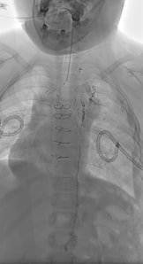

Indications

  • Postoperative lymphatic leakage (chyle leak, chylothorax, chylous ascites)
  • High-output chylothorax (>1000 mL/day) refractory to conservative management
  • Traumatic chylothorax following thoracic procedures
  • Central conducting lymphatic anomaly (CCLA), kaposiform lymphangiomatosis (KLA)
  • Plastic bronchitis with lymphatic etiology
  • Protein-losing enteropathy due to intestinal lymphatic dysfunction

Contraindication

  • None absolute.
  • Severe cardiopulmonary disease (then use water-soluble contrast)
  • Known right-to-left cardiac shunt (then use water-soluble contrast)
  • Active pulmonary infection or severe respiratory compromise
  • Severe coagulopathy (INR >2.0, platelets <50,000/mm³), only if thoracic duct embolization planned
  • Pregnancy (radiation exposure concerns)

Pre-procedure

Essential Laboratory Studies
  • Complete blood count with platelet count
  • Coagulation studies (PT/INR, aPTT)
  • Comprehensive metabolic panel (renal function assessment)
  • Liver function tests (albumin, protein levels)
​Imaging Requirements
  • Chest X-ray or CT chest (baseline pulmonary status)
  • Cross-sectional imaging (CT/MRI) to identify lymphatic pathology
  • Ultrasound evaluation of inguinal lymph nodes (pre-procedure planning)
  • Echocardiogram if cardiac shunt suspected
Additional Assessments
  • Pulmonary function tests in patients with respiratory disease
  • Drain fluid analysis (triglycerides, chylomicrons, cell count)
  • Nutritional assessment (albumin, pre-albumin, lymphocyte count)
​Antibiotics
Only if embolization is considered
  • Cefazolin and metronidazole
  • Alternative clindamycin and gentamicin


Technique

Supplies
  • Needles: 22-25G hypodermic needles.
  • Short connecting tubes, 3-5 ml syringes.
  • Lipiodol dose: 0.25 ml/kg, not to exceed 10 ml.
  • If embolizing, 70 cm Rapid Transit microcatheter, V18 guidewire, microcoils (e.g. Embold coils), histoacryl glue (1:1 mixture with lipiodol).
 
Prep
  • Bilateral inguinal regions and upper thighs are prepped. The left upper chest and neck are also prepped to allow sonographic evaluation of the terminal portion of the thoracic duct. If there is potential for cannulation of the thoracic duct, the entire abdomen should be prepped
 
Technique
  • Connect needle via two short connecting tubes to 3 ml syringe with lipiodol
  • Initial snapshot images to cover from neck to upper thighs.
7 year old boy with CCLA. Intranodal lymphangiogram demonstrates delayed progression of contrast with reflux and dilated terminal portion of the thoracic duct.
  • Using a high-frequency transducer, target bilateral inguinal lymph nodes.  Advance needle to the corticomedullary junction. Avoid through and through puncture.
  • Stabilize the needles with Steri‐Strip and avoid manipulation of needles once inserted into node.
  • Inject lipiodol at a slow rate to avoid extravasation. Average 1 ml every 5-10 minutes.
  • Real-time intermittent fluoroscopic imaging.
  • Serial spot imaging every 5-10 minutes.
  • Flush with saline. Abdominal massage can be performed if stasis within the abdomen.
  • Progressive upward imaging from pelvis to chest
  • Documentation of lymphatic anatomy and pathology
  • Identification of leak sites or obstruction
  • Assessment for collateral pathways

Complications

Immediate Complications (0-24 hours)
  • Contrast extravasation (5-15% incidence)
    • Usually asymptomatic
    • Local pain or swelling at injection site
    • Rarely requires intervention
  • Pulmonary oil embolism (dose-dependent risk)
    • Risk increases with volume >18 mL
    • Usually asymptomatic or mild dyspnea
    • Managed with supportive care and oxygen
  • Lymph node injury or bleeding
    • Minor hematoma formation
    • Rarely clinically significant
    • Managed with compression and observation
Early Complications (24-72 hours)
  • Infection at puncture site (rare <1%)
    • Local cellulitis or lymphangitis
    • Antibiotic therapy usually sufficient
    • Rarely progresses to systemic infection
  • Delayed hypersensitivity to ethiodized oil
    • Rare allergic reactions
    • Managed with antihistamines and steroids
Late Complications (>72 hours)
  • Pulmonary complications (volume-dependent)
    • Transient pulmonary infiltrates
    • Usually resolve spontaneously
    • May require corticosteroid therapy in severe cases
  • Failed therapeutic effect (20-30% of cases)
    • Persistent lymphatic leakage
    • May require repeat procedure or alternative intervention
    • Consider thoracic duct embolization

Thoracic Duct Embolization


Technique

  • Angle image identifier at 15-20 degrees craniocaudal to provide an end-on view.
  • Identify cisterna chyli
  • 21-22 gauge Chiba needle advanced into the cisterna chyli or another targeted duct.
  • A 5-10 degree curve on the needle can sometimes be helpful in gaining access.
  • Consider pre-loading with guidewire.
  • If difficulty accessing try different levels (sometimes maceration of thoracic duct may be enough to terminate leak).
  • Advance guidewire (typically V18, (Boston Scientific, Natick, MA) ) to cranial portion of thoracic duct to ensure secure access.
  • Exchange needle for microcatheter (most commonly Rapid Transit)
  • If leak identified, 1-2 micro-coils can be placed for extra safety
  • Follow with injection of histoacryl (1;1 with lipiodol). Further dilution risks venous embolization as polymerization in chyle is slower than in blood.
  • Withdraw catheter rapidly while aspirating.

Complications

Immediate Complications (0-24 hours)
  • Access site bleeding (2-5% incidence)
    • Retroperitoneal hematoma (rare but serious)
    • Managed with observation, blood products if needed
    • CT imaging for large hematomas
  • Bowel injury (<1% incidence)
    • Perforation during transabdominal access
    • Recognition by contrast extravasation
    • Surgical consultation and repair if needed
  • Vascular injury (rare)
    • Aortic or IVC puncture
    • Immediate recognition and management
    • Vascular surgery consultation

Early Complications (24-72 hours)
  • Infection (1-3% incidence)
    • Access site infection or systemic sepsis
    • Broad-spectrum antibiotic therapy
    • Drainage if abscess formation
  • Contrast reaction (rare)
    • Allergic reaction to ethiodized oil
    • Standard allergic reaction management
    • Corticosteroids and supportive care

Procedure-Related Complications
  • Failed access (10-20% incidence)
    • Inability to catheterize thoracic duct
    • Consider alternative approach or thoracic duct disruption
    • May require repeat procedure
  • Glue migration (rare)
    • Embolization of non-target vessels
    • Usually clinically insignificant
    • Monitor for systemic effects

​Long-Term Complications (>1 month)
  • Chronic diarrhea (reported in 10-15% of patients)
    • May be related to lymphatic disruption
    • Usually mild and manageable
    • Part of informed consent discussion
  • Lower extremity edema (5-10% incidence)
    • Secondary to altered lymphatic drainage
    • Managed with compression therapy
    • Usually improves over time
  • Contralateral chylothorax (rare)
    • Due to cisterna chyli access point leakage
    • Prevented by accessing via lumbar ducts
    • May require repeat embolization
  • Nutritional complications (uncommon)
    • Fat-soluble vitamin deficiency
    • Protein malnutrition in severe cases
    • Nutritional monitoring and supplementation

References

  • Itkin M, Kucharczuk JC, Kwak A, et al. Nonoperative thoracic duct embolization for traumatic thoracic duct leak: experience in 109 patients. J Thorac Cardiovasc Surg. 2010;139(3):584-589.
  • Nadolski GJ, Itkin M. Feasibility of ultrasound-guided intranodal lymphangiography in children. Pediatr Radiol. 2012;42(9):1057-1061.
  • Pieper CC, Hur S, Sommer CM, et al. Back to the future: lipiodol in lymphography-from imaging to treatment. Invest Radiol. 2019;54(9):600-615.
  • Baş A, Üstündağ A, Özdemir M, et al. Efficiency of intranodal lymphangiography in the treatment of postoperative lymphatic leakage. Ulus Travma Acil Cerrahi Derg. 2025;31(3):221-225.
  • Chick JF, Reddy SN, Jeffers AB, et al. Intranodal lymphangiography with water-soluble iodinated contrast medium for imaging of the central lymphatic system. Radiology. 2022;302(2):431-439.
  • Jardinet T, Veer HV, Nafteux P, et al. Intranodal lymphangiography with high-dose ethiodized oil shows efficient results in patients with refractory, high-output postsurgical chylothorax: a retrospective study. AJR Am J Roentgenol. 2021;217(2):433-438.
  • Maleux G, Indesteege I, Laenen A, et al. Complications during lymphangiography and lymphatic interventions. Tech Vasc Interv Radiol. 2020;23(3):100688.
  • Lee EW, Shin JH, Ko HK, et al. Lymphangiography to treat postoperative lymphatic leakage: a technical review. Korean J Radiol. 2014;15(6):724-732.
  • Sommer CM, Pieper CC, Itkin M, et al. Conventional lymphangiography (CL) in the management of postoperative lymphatic leakage (PLL): a systematic review. Rofo. 2020;192(11):1025-1035.
  • Gupta A, Naranje P, Vora Z, et al. Intranodal lipiodol injection for the treatment of chyle leak in children - a preliminary experience. Br J Radiol. 2022;95(1137):20211270.
  • Chaudry G. Complex Lymphatic Anomalies and Therapeutic Options. Tech Vasc Interv Radiol. 2019;22(4):100632.

Home

Essentials

Protocols

Procedures

Online Library

about us

Copyright © Pediatric IR Handbook 2025
  • Home
  • Procedures
  • Protocols
  • Online Library
    • Pediatric IR Papers
    • Presentations and Webinars
    • IR Equipment and IFU
  • BCH IR Fellows Homepage
  • About Us